Is there a risk of nerve damage during wisdom teeth surgery?
- 5 days ago
- 6 min read

Numbness after a dental procedure is expected — for a few hours. But when that numbness, tingling, or burning sensation in the lip, chin, cheek, or tongue lasts for days, weeks, or longer, it has a name: paresthesia.
It is one of the complications patients worry about most before wisdom tooth surgery, and understandably so — the lips and tongue are central to eating, speaking, and simply feeling like yourself.
Dr. Zach Saltman, board-certified oral & maxillofacial surgeon and founder of Saltman Implantology & Oral Surgery, hears this concern and addresses it directly during every pre-surgical evaluation.
This article explains what paresthesia actually is, why it happens, how Dr. Saltman assesses and manages nerve-related risk, and — most importantly — how modern surgical planning dramatically reduces that risk.
What Paresthesia Actually Is
Paresthesia is an altered sensation — numbness, tingling, prickling, or a burning "pins and needles" feeling — caused by irritation or injury to a sensory nerve. In dentistry, it most often affects the lower lip, chin, or tongue after lower wisdom tooth removal, or occasionally after a local anesthetic injection.
It's important to distinguish paresthesia from the normal numbness caused by local anesthesia, which wears off within a few hours. Paresthesia is diagnosed when altered sensation persists well beyond the expected duration of the anesthetic — typically defined as lasting more than 24 hours, and in the case of prolonged or permanent injury, months or even years.
The Nerves at Risk
Two nerves are most commonly involved in lower jaw procedures.
The inferior alveolar nerve (IAN) runs through a bony canal inside the lower jaw, supplying sensation to the lower teeth, lip, and chin.
The lingual nerve runs along the inner surface of the jawbone, close to the gum tissue near the wisdom tooth site, and supplies sensation and taste to the front two-thirds of the tongue.
The anatomic relationship between a lower wisdom tooth's roots and the inferior alveolar canal varies enormously from person to person. In some patients the roots sit well above the canal with a comfortable margin of bone between them; in others, the roots wrap around the canal, sit directly against it, or even pass through it. This variability is exactly why Dr. Saltman relies so heavily on 3D imaging before recommending an extraction — a topic we return to below.
How Nerve Injury Happens
Surgical injury to the IAN typically results from the close proximity of the wisdom tooth roots to the nerve canal. During extraction, root removal, bone removal (osteotomy), or the use of elevators to loosen a tooth can stretch, compress, bruise, or in rare cases directly injure the nerve. The vast majority of these injuries are due to compression, stretching, or localized inflammation — not the nerve being cut.
Injection-related injury is a separate mechanism, most often implicating the lingual nerve. It can occur if a needle directly contacts a nerve fascicle during a nerve block injection (patients sometimes describe a brief electric "zing" sensation at the moment of injection), or if the anesthetic solution itself causes localized chemical irritation within the nerve sheath. A small hematoma (blood collection) forming near the nerve can also create temporary compression.
Who Is at Higher Risk
Certain factors increase the statistical likelihood of nerve involvement, and Dr. Saltman discusses these with every patient individually before surgery:
Deeply or horizontally impacted lower wisdom teeth, particularly those whose roots appear on imaging to be superimposed on, in contact with, or wrapping around the mandibular canal.
Increasing patient age, since roots are more fully formed and bone is denser, which can require more force during extraction.
Complex root anatomy — curved, divergent, or unusually long roots.
A history of a difficult or prolonged surgical extraction.
For injection-related injury: individual anatomic variation in nerve position, which cannot always be predicted in advance.
Classifying Nerve Injuries
Oral surgeons often describe nerve injuries using the Seddon classification, which is useful for understanding prognosis:
Neurapraxia — the mildest form. The nerve fiber itself is intact but temporarily unable to conduct signals, usually due to compression or stretching. Sensation typically returns within days to a few weeks.
Axonotmesis — a moderate injury in which the nerve fiber (axon) is damaged but its surrounding support structure remains intact. Because nerves regenerate slowly — roughly one millimeter per day — recovery can take several months.
Neurotmesis — the most severe injury, involving complete disruption of the nerve. Meaningful spontaneous recovery is unlikely, and microsurgical repair may be the only path to regaining sensation.
The encouraging reality is that the overwhelming majority of paresthesia cases following wisdom tooth extraction fall into the neurapraxia or mild axonotmesis category and resolve on their own.
Symptoms to Watch For
Patients with nerve involvement may notice: numbness of the lip, chin, cheek, or tongue; a tingling or "asleep" sensation that doesn't fade; altered or absent taste sensation on one side of the tongue; drooling on the affected side without realizing it; accidentally biting the lip, cheek, or tongue because sensation is dulled; and, when the lingual nerve is involved, subtle changes in speech clarity or difficulty forming certain sounds.
How Nerve Injury Is Diagnosed and Monitored
If paresthesia is suspected, Dr. Saltman will perform serial neurosensory testing — assessing the patient's response to light touch, pinprick sensation, two-point discrimination, and temperature — and document these findings at follow-up visits to track whether sensation is returning over time.
In select cases, a cone-beam CT (CBCT) scan is used to evaluate the position of any retained root fragments, hardware, or bone relative to the nerve canal.
If paresthesia is suspected, Dr. Saltman and his team perform serial neurosensory testing — assessing the patient's response to light touch, pinprick sensation, two-point discrimination, and temperature — and document these findings at follow-up visits to track whether sensation is returning over time.
Treatment and Management Options
Management is tailored to the type and severity of the injury:
Observation. Because most cases are neurapraxia, watchful waiting with scheduled sensory re-testing is the appropriate first step for mild cases.
Anti-inflammatory medication. In the early post-injury period, corticosteroids may be prescribed to reduce swelling and inflammation around the nerve, which can shorten recovery time.
Vitamin B complex supplementation. Some surgeons recommend B-vitamin supplementation to support nerve tissue health during recovery, though evidence for this is supportive rather than definitive.
Neurosensory re-education. Structured exercises that stimulate the affected area can help the brain "remap" sensation as nerve fibers regenerate.
Microsurgical nerve repair. If there is no measurable improvement by around three months, and imaging or clinical findings suggest a significant injury, referral to a microsurgeon for nerve exploration and repair may be recommended.
Outcomes are generally best when repair is performed within six to twelve months of the original injury, since nerve and muscle tissue become progressively less receptive to reinnervation over time.
Prevention: Why Imaging and Planning Matter So Much
The single most effective way to reduce the risk of nerve injury is thorough preoperative evaluation. A standard panoramic X-ray gives a two-dimensional view and can suggest that roots are close to the canal, but it cannot always show the true three-dimensional relationship. When a panoramic film raises concern, a CBCT scan provides a three-dimensional view that lets Dr. Saltman see exactly how the roots relate to the nerve canal from every angle.
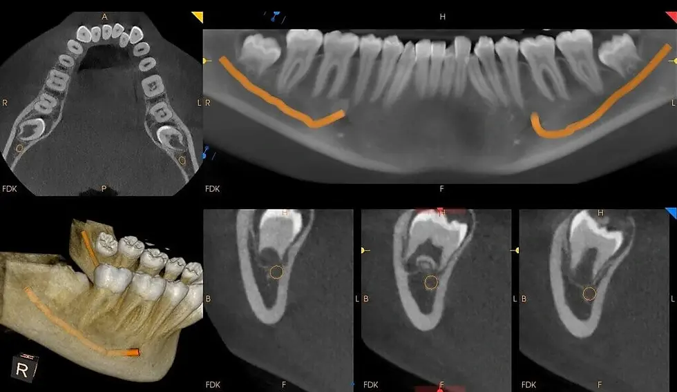
In cases where imaging shows a high-risk relationship, Dr. Saltman has options beyond a standard extraction. Saltman Oral Surgery offers coronectomy when appropriate — a technique in which the crown of the tooth is removed but the roots are intentionally left in place to avoid disturbing the nerve — as well as modified surgical approaches that minimize manipulation near the canal. Dr. Saltman discusses these options, along with each patient's individual risk profile, as part of an informed consent conversation before any wisdom tooth surgery.
The Bottom Line on Nerve Damage Risk in Wisdom Teeth Removal
Permanent nerve injury after wisdom tooth extraction is uncommon, and temporary paresthesia — when it does occur — resolves in the large majority of cases without intervention. What matters most is choosing a surgical team that takes 3D imaging seriously, explains your individual risk factors clearly, and monitors you closely if any altered sensation does occur.
At Saltman Oral Surgery, every wisdom tooth case involving teeth close to the nerve canal is evaluated by Dr. Saltman with appropriate 3D imaging before a treatment plan is finalized. When imaging suggests elevated risk, Dr. Saltman will discuss whether coronectomy or a modified surgical approach is the right choice for you. Any patient who experiences post-operative numbness is followed with structured sensory testing until resolution.
Have Questions About Your Own Risk Factors?
If you're concerned about nerve involvement before an upcoming wisdom tooth surgery, or you're experiencing altered sensation after a procedure, schedule a consultation with Dr. Saltman — early evaluation gives you the best chance of a straightforward recovery.
Saltman Implantology & Oral Surgery
📍 Washington, DC: 1426 21st Street NW, Second Floor, Washington, DC 20036
📍 McLean, VA: 1363 Beverly Road, Suite 250, McLean, VA 22101


Comments